Why Seamless IMS-LIS Integration Is Essential for Digital Pathology
Unified digital workflows connecting IMS to LIS unlock efficiency, compliance, and the full potential of AI for modern pathology
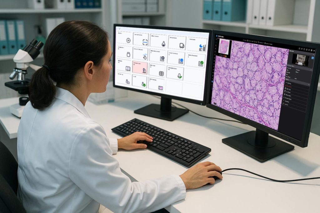
Digital pathology has moved decisively from experimentation into everyday practice. For pathology organizations, it now underpins clinical workflows, operational efficiency, and long-term strategy. To enable remote sign-out and AI-driven analysis, organizations have turned to Image Management Systems (IMS) that can organize, track, and contextualize whole slide images across the diagnostic workflow.
However, the data needed to interpret those images extends well beyond what an IMS typically manages. Patient demographics, clinical context, specimen metadata, test orders, results, finalized reports and more all live in a separate but equally critical system: the Laboratory Information System (LIS). For groups accustomed to thinking primarily in terms of image workflows, the LIS can feel like a parallel universe, yet it is the system of record for nearly all non-image pathology data. When IMS and LIS platforms operate in isolation, inefficiencies emerge, context is lost, and the promise of digital pathology remains only partially realized.
As digital pathology programs mature, it is becoming evident that success depends not only on performance, scale, and regulatory readiness, but also on seamless interoperability. In this article, we explore why deep, context-aware integration between an IMS and a modern LIS is essential to unlocking the full value of digital pathology. We’ll examine the practical challenges this integration resolves and the strategic benefits it enables for pathology groups advancing toward a fully digital, AI-enabled future.
Image Management Systems (IMS) in Digital Pathology
A digital pathology image management system (IMS) goes far beyond mere image viewing. At its best, it is an enterprise-grade platform designed to manage the entire lifecycle of digital pathology images across clinical, research, and educational workflows, and serving as the backbone of digital pathology operations, supporting everything from secure storage to AI integration and multi-institution collaboration.
Core Capabilities of a Modern Digital Pathology IMS
A robust IMS typically includes:
- Advanced Slide Viewing: High-performance visualization across multiple formats (SVS, NDPI, SCN, DICOM, BigTIFF, and more), synchronized multi-slide viewing, annotations, overlays, and fast rendering.
- Centralized Data Management: Secure, scalable image storage, on-premises, cloud, or hybrid, with governance controls and long-term data stewardship.
- Security and Compliance: HIPAA-grade protections, encryption, audit trails, role-based access, and regulatory readiness for clinical use.
- Workflow Integration: Case management, task tracking, and seamless interoperability with LIS, EHR, PACS, and other enterprise laboratory software systems.
- User Roles and Access Control: Granular permissions ensure that only authorized users access sensitive data, while maintaining accountability.
- Collaboration Tools: Support for remote consults, tumor boards, education, and peer review through shared environments.
- AI and Image Analysis Enablement: Integration with AI models for detection, quantification, and pattern recognition, embedded directly into diagnostic workflows.
- Regulatory Readiness: Many IMS platforms are designed to meet FDA, HIPAA, GDPR, IVDR, and other regulatory standards. Some, such as PathPresenter’s clinical viewer, are FDA 510(k) cleared and EU-IVDR certified for primary diagnosis when paired with approved scanners.
In short, an IMS provides a comprehensive infrastructure for digital pathology. It can be highly capable on its own, handling slide storage, visualization, annotations, and even AI outputs. But when it operates independently from the laboratory’s core operational system, its impact is inherently limited. Rather than dissolving legacy barriers, a disconnected IMS can unintentionally recreate them in digital form.
True digital pathology maturity is reached only when images and laboratory data function as a single, coherent workflow. Without that alignment, pathology groups often find themselves managing parallel systems that never quite converge.
The Operational Cost of Keeping IMS and LIS Separate
When image workflows and laboratory workflows are not connected at a foundational level, pathology teams encounter familiar but amplified challenges, including:
- Reliance on manual reconciliation of cases and slides
- Repeated entry of patient and specimen data
- Frequent toggling between systems during case review
- Greater exposure to mismatches, omissions, and reporting errors
- Disjointed compliance records spanning multiple platforms
- Collaboration slowdowns across sites and subspecialties
In isolation, each issue may seem manageable. At scale however, particularly in regulated, high-throughput environments, they compound rapidly, eroding both efficiency and confidence in the digital workflow.
Rethinking the LIS: More Than a Back-End Database
For many digital pathology teams, the LIS is perceived primarily as a static repository: the place where orders originate and reports ultimately land. While that historical role remains essential, it no longer reflects how modern laboratories operate.
From Passive Record-Keeping to Active Workflow Orchestration
Today’s laboratories increasingly require systems that do more than store information. A modern LIS such as LigoLab functions as an operational engine: driving workflow logic, enforcing rules, automating handoffs, and presenting the right context at the right moment.
When an IMS is natively integrated into this environment, digital pathology is no longer an external tool that users “visit.” Instead, image review becomes a natural extension of the laboratory workflow, governed by the same logic, permissions, and traceability as the rest of the case lifecycle.
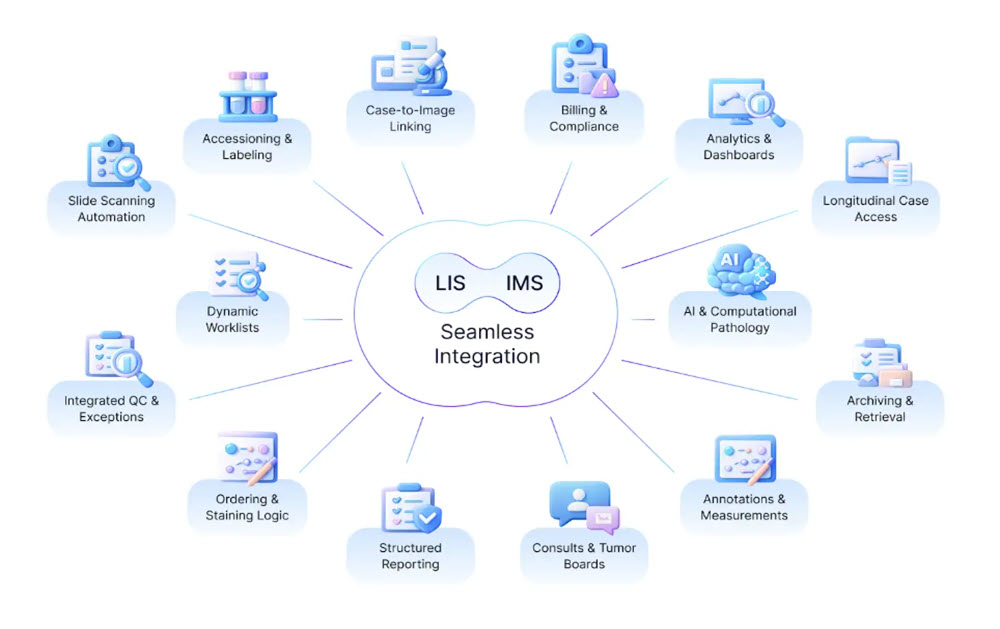
What Integrated LIS–IMS Workflows Enable Day to Day
In a genuinely unified digital pathology environment:
- Digital slides are opened directly from the laboratory case, not searched for separately
- Patient, specimen, and order context flows automatically into image review
- Movement between data, images, and reports feels continuous rather than segmented
- Auditability covers both diagnostic decisions and image interactions end to end
- AI outputs appear within established workflows rather than as side-channel tools
This level of integration changes how work is performed: not by adding features, but by removing friction.
Core Challenges Solved by Deep Integration
1. Smoother, Faster Diagnostic Workflows
When image access is embedded within the laboratory system, pathologists spend less time navigating software and more time interpreting cases. Reduced friction translates directly into faster turnaround and lower cognitive load.
2. Scalable Support for Remote Practice
Integrated platforms make location largely irrelevant. Secure remote sign-out, real-time collaboration, and distributed subspecialty review become standard operations rather than exceptions.
3. Better Decisions Through Complete Context
Images rarely tell the full story on their own. Tight coupling between LIS and IMS ensures that diagnostic interpretation always occurs alongside the complete clinical and specimen context, reducing the risk of incomplete assessment.
4. Stronger Governance and Compliance
When images and laboratory data share a unified audit framework, compliance with regulatory and accreditation requirements becomes simpler and more defensible—without added administrative burden.
5. A Practical Path to Enterprise AI
AI delivers value only when it operates inside real workflows. Integrated LIS–IMS environments provide the governance, data access, and workflow hooks required to deploy AI responsibly and at scale.
Integration in the Real World: From Concept to Practice
A practical example of this approach can be seen in the collaboration between PathPresenter and LigoLab. Rather than treating image management as an external system, PathPresenter’s clinical viewer is embedded directly within LigoLab’s enterprise laboratory platform.
The result is a unified diagnostic experience where pathologists interact with slides in the same environment where cases are managed, orders are tracked, and reports are finalized. This tight coupling reduces navigation overhead, accelerates review cycles, and establishes a solid foundation for future AI-driven enhancements.
Building Toward a Unified Digital Pathology Future
At its core, LIS–IMS integration is not about software convenience. It is about redefining how pathology work flows from accessioning through diagnosis and reporting.
When laboratory data and images operate as one system:
- Operations gain visibility and automation
- Financial teams benefit from cleaner alignment with billing and revenue workflows
- Clinical staff gain modern tools that reduce manual effort
- Patients benefit from faster, more consistent diagnostic outcomes
As digital pathology, AI, and remote diagnostics continue to evolve, this alignment moves from “nice to have” to mission critical. Pathology groups that invest early in deep, contextual integration position themselves not just to digitize slides, but to fundamentally modernize how pathology is practiced.
More Information
Looking for a new (or improved) LIS-IMS integration? Connect with our team to assess your workflow needs and explore the best solution.